 |
Brilliant Medical System Pte Ltd | |||||||
| Your Trusted Medical Equipment Supplier | ||||||||
|
||||||||
Product Category
|
Software |
Product>SoftwareAMCAD
|
||||
|---|---|---|---|---|
AmCAD Software
|
||||
AmCAD-UT® Detection uses statistical pattern recognition and quantification methods to perform analytical processing of images. The physician may process the image for detection of sonographic characteristics (i.e., hyperechoic foci, echogenicity, texture, margin, anechoic areas, taller than wide, tumor shape, and tumor size) with assistance of AmCAD-UT® Detection.AmCAD-UT® Detection provides more detailed information with quantification and visualization of the sonographic characteristics on thyroid nodule that may assist medical professionals in making their diagnostic decision. |
||||
| Detection process of AmCAD | ||||
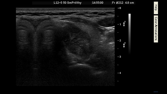 |
Open Ultrasound image saved before. |
|||
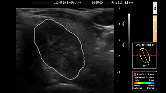 |
Define nodule segmentation |
|||
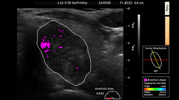 |
Detect the Anechoic area |
|||
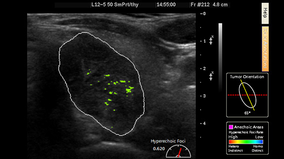 |
Show the Hyperrechoic Foci |
|||
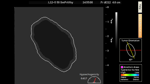 |
Display Echogenicity with contrast, show the possibllity of malignancy via "taller-than-wide" shape. |
|||
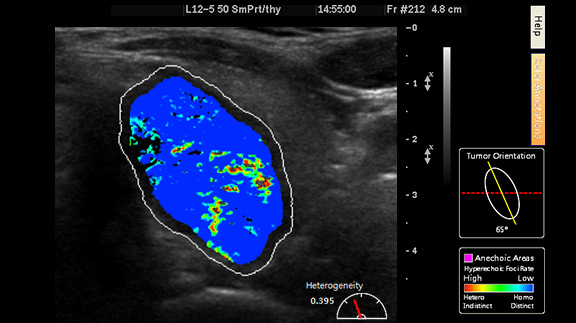 |
Display the texture, show the possibllity of malignancy via "taller-than-wide" shape. |
|||
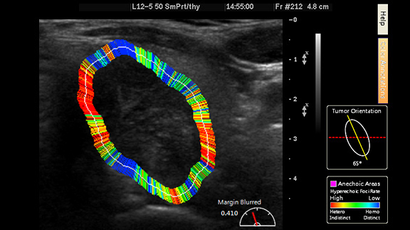 |
Show the Margin, show the possibllity of malignancy via "taller-than-wide" shape. |
|||
| According to survey results, more than 300 million people around the world suffer from thyroid disease. The prevalence for women is 8 times higher than men. The incidence rate for women aged 40 or above is 10% or higher. Therefore, thyroid cancer has become one of the most concerning and troubling cancers in the world. | ||||
| Different physicians may arrive at different interpretations over the same image; sometimes even the same physician may conclude different interpretations over the same image at different times. The inconsistency of the results may reach up to 70%. | ||||
| Even though physicians who made a definitive diagnosis and thus performed thyroidectomy, still up to 50% of results turned out to be benign. | ||||
| The “AmCAD-UT Detection” utilizes patented image analysis technology to capture commonly used clinical diagnostic features as well as the pattern features of the thyroid neoplasm from the thyroid ultrasound images. Through quantification and visualization, detailed color images are thereby displayed for greatly improved and more accurate ultrasound image interpretation. With the ever increasing global incidence of thyroid cancer, such technology provides a non-invasive assessment tool for the definitive diagnosis of thyroid cancer. | ||||
|
|
||||
|
||||